
Tanilba Bay Vet
Vet Radiology in Tanilba Bay
- Pet Grooming
- Pet Health Packages
- Surgical Care
Introduction
Radiology & Ultrasonography for Pets
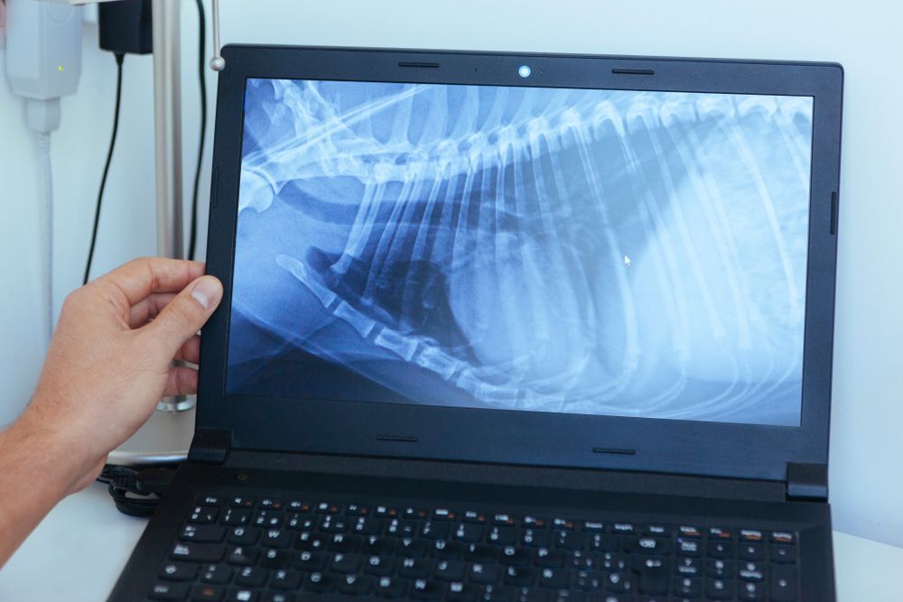
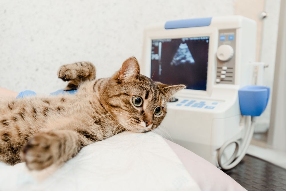
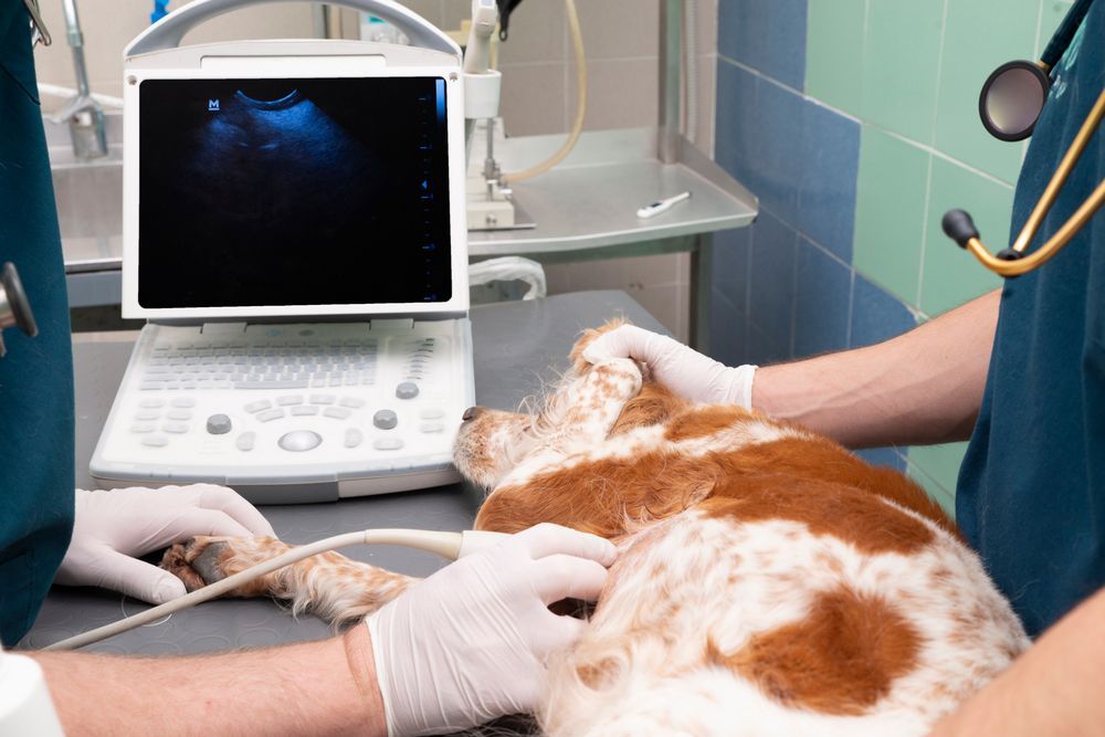
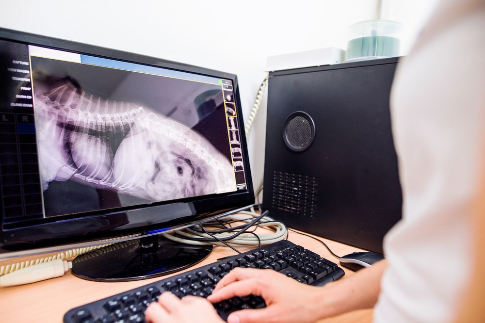
When pets develop health issues that are not visible from the outside, imaging becomes an effective diagnostic tool. Vet radiology and ultrasonography allow veterinarians to see inside the body without invasive procedures, giving valuable insights into organs, bones and soft tissues. These methods are particularly helpful for investigating persistent illness, injuries or unexplained symptoms. X-rays are commonly used to examine skeletal structures, while ultrasound provides live images of internal organs.
At Tanilba Bay Vet, our modern imaging equipment supports accurate assessments that guide decisions about care. These services are often combined with other diagnostic methods to create a fuller picture of health.
For safe imaging and compassionate support, call Tanilba Bay Vet today on (02) 4024 2004.
Submit an Enquiry
Thank you for contacting Tanilba Bay Vet.
We will get back to you as soon as possible.
Oops, there was an error sending your message.
Please try again later.
Our Service
Common Uses of Veterinary Imaging
Radiology and ultrasonography can be applied in many situations to assist with diagnosis and treatment planning:
- Bone & Joint Assessment
X-rays are valuable for identifying fractures, joint issues or signs of arthritis, providing images that assist with treatment decisions. - Abdominal Health
X-rays and ultrasound helps evaluate organs like the liver, kidneys and bladder, detecting changes such as stones, inflammation or blockages. - Foreign Object Detection
Both X-rays and ultrasound can reveal ingested items or obstructions, guiding the safest way to address the problem.
These applications highlight the versatility of imaging in veterinary medicine.
Need To Know
The Value of Imaging in Veterinary Care
Imaging is not always the first step in diagnosis, but it is often recommended when physical examinations and laboratory tests do not provide enough answers. Radiology and ultrasonography allow conditions to be investigated in greater detail, giving insight into the extent or location of a problem. These results are interpreted by qualified veterinarians who consider the pet’s history, behaviour and overall health.
In some cases, imaging may highlight issues that require further testing or specialist referral. Owners can take comfort knowing that these tools are non-invasive and designed to minimise stress for animals. By providing clearer information, imaging helps guide safer and more effective care plans for pets of all ages.
Need Help?
Frequently Asked Questions
When might my pet need an X-ray?
X-rays are usually recommended when there are concerns about bones, joints or internal structures that cannot be examined externally. They can detect fractures, arthritis or swallowed objects. Sometimes X-rays are also used to check lung health or heart size. Your veterinarian will only recommend imaging when it provides meaningful information for your pet’s care.
How does ultrasound differ from X-rays?
X-rays capture still images of dense tissues like bones and lungs, making them useful for structural issues. Ultrasound uses sound waves to show soft tissue organs in real time. This makes it effective for examining the heart, liver, kidneys and bladder. The two methods are often used together to provide a clearer overall picture.
Is imaging safe for pets?
Yes, both X-rays and ultrasound are considered safe when performed by trained professionals. X-rays involve low doses of radiation, and protective measures are used to minimise exposure. Ultrasound uses sound waves, which are non-invasive and painless. These techniques are widely used because they provide valuable information without significant risk.


